2026
Morgan C, O'Malley O, Lüleci E, Saraogi C, Reilly P, Kedgley AE. (2026) Get a Grip: How Hand Anthropometrics, Grip Strength, and Sex Influence Power Tool Use in Orthopaedics. JB & JS Open Access. doi: 10.2106/JBJS.OA.25.00343
Schweizer A, Horwitz MD, Kedgley AE, Honigmann P. (2026) Structural balance restoration in complex forearm problems using 3D technology. The Journal of Hand Surgery, European Volume doi: 10.1177/17531934261430112
Lüleci EH, Kedgley AE. (2026) Evaluating the Effectiveness of Age Simulation Gloves. Human Factors doi: 10.1177/00187208261434691
Han Y, Vallerini FM, Holdsworth F, Wanglertpanich K, Kedgley AE, Masouros SD. (2026) A custom force plate for quantifying the force applied by the finger during smartphone usage. Frontiers in Bioengineering and Biotechnology doi: 10.3389/fbioe.2026.1685410
Raftery KA, Levy H, Singh R, Madi M, Slater TD, Crossman AJ, Kedgley AE, Freedman BA, Newell N. (2026) Intervertebral disc distraction stiffness predicts endplate subsidence following transforaminal interbody cage expansion: an ex vivo study, European Spine Journal doi: 10.1007/s00586-025-09715-x
2025
Crossman AJ, Kedgley AE. (2025) A novel approach to muscle control for in vitro gait simulators, Annual International Conference of the IEEE Engineering in Medicine and Biology Society doi: 10.1109/EMBC58623.2025.11252873
Devanand DB, Gardiner MD, Kedgley AE. (2025) A compact orthosis compliance monitoring device using pressure sensors and accelerometers: design and proof-of-concept testing, Sensors doi: 10.3390/s25051352
Miller R, Kedgley AE, Farnebo S, Stockmans F, Zlotolow DA, Horwitz MD. (2025) Round table discussion. Integration of artificial intelligence into daily practice, Journal of Hand Surgery (European Volume), doi: 10.1177/17531934251342418
2024
Gracia-Ibáñez LV, Mohseni M, Kedgley AE, Jarque-Bou NJ, Granell P, Vergara M, Sancho-Bru JL. (2024) Electromyography parameters to discriminate hand osteoarthritis and infer their functional impact, Sensors doi: 10.3390/s24206706
Katakura M, Rezende MAG, Calder JDF, Kedgley AE. (2024) A comparison of abductor hallucis muscle activation and medial longitudinal arch angle during nine different foot exercises, Gait & Posture, doi: 10.1016/j.gaitpost.2024.06.008
Sehjal R, Rusli W, Kedgley AE, Sagmeister ML, Williamson M, Smith A. (2024) Biomechanical comparison of 5 different fixation constructs in a trapeziometacarpal joint arthrodesis model, The Journal of Hand Surgery (American Volume), doi: 10.1016/j.jhsa.2022.12.010
Watson FCE, Kedgley AE, Schofield S, Behan FP, Boos CJ, Fear NT, Bennett AN, Bull AMJ. (2024) Upper limb function in people with upper and lower limb loss 8 years postinjury: The Armed Services Trauma Outcome Study (ADVANCE) Cohort Study. Physical Therapy. doi: 10.1093/ptj/pzae082
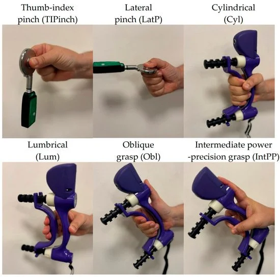
Recording maximal voluntary force in six grasp types relevant for functionality.

Exercises to strengthen intrinsic foot muscles, include toe curl exercises, short-foot exercises, unilateral balance activities and toe spread exercises.

Five fixation methods used for trapeziometacarpal joint arthrodesis.
2023
Devanand DB, Kedgley AE (2023) Objective methods of monitoring usage of orthotic devices for the extremities: A systematic review, Sensors, doi: 10.3390/s23177420
Katakura M, Kedgley A, Shaw J, Mattiussi A, Kelly S, Clark R, Allen N, Calder J. (2023) Epidemiological characteristics of foot and ankle injuries in 2 professional ballet companies: a 3-season cohort study of 588 medical attention injuries and 255 time-loss injuries, Orthopaedic Journal of Sports Medicine, doi: 10.1177/23259671221134131
Li LX , Kedgley AE, Horwitz MD. (2023) A Review of the use of 3D printing technology in treatment of scaphoid fractures, The Journal of Hand Surgery (Asian-Pacific Volume) doi: 10.1142/S2424835523500042
McMenemy L, Mondini V, Roberts DC, Kedgley A, Clasper JC, Stapley SA. (2023) Pattern of upper limb amputation associated with lower limb amputation: the UK military experience from Iraq and Afghanistan. BMJ Military Health. doi: 10.1136/bmjmilitary-2021-001783
Wai G, Rusli WMR, Ghouse S, Kieser DC, Kedgley AE, Newell N. (2023) Statistical shape modelling of the thoracic spine for the development of pedicle screw insertion guides, Biomechanics & Modeling in Mechanobiology, doi: 10.1007/s10237-022-01636-8

Mechanisms of medical-attention (MA) and time-loss (TL) injuries in female and male dancers by injury type. The 5 most common types of injuries are shown.

Distance maps representing the morphological variations in thoracic vertebrae (T4–T6) along the first three principal components − 3 standard deviations (SD) from the mean (left column) and + 3SD from the mean (right column)

Level of amputation by anatomical region.
2022
Gionfrida L, Rusli WMR, Kedgley AE, Bharath AA. (2022) A 3DCNN-LSTM multi-class temporal segmentation for hand gesture recognition, Electronics, doi: 10.3390/electronics11152427
Chung VWJ, Newell R, Kedgley A, Anglin C, Masri BA, Hodgson AJ. (2022) Verifying a C-arm-based roentgen stereophotogrammetric analysis protocol for assessing tibial implant movement in total knee arthroplasty, Medical & Biological Engineering & Computing, doi: 10.1007/s11517-022-02594-0
Jones D, Vardakastani V, Kedgley AE, Gardiner MD, Vincent TL, Culmer PR, Alazmani A. (2022) HAILO: A sensorised hand splint for the exploration of interface forces. IEEE Transactions on Biomedical Engineering. doi: 10.1109/TBME.2022.3155589
Vissers G, Rusli WMR, Scarborough A, Horwitz MD, McArthur GJ, Kedgley AE. (2022) A study to compare strengths of cadaveric tendon repairs with round-bodied and cutting needles, Journal of Hand Surgery (European Volume), doi: 10.1177/17531934211064201
Shaerf DA, Chae WJ, Sharif-Razavian R, Vardakastani V, Kedgley AE, Horwitz, M. D. (2022). Do “anatomic” distal ulna plating systems fit the distal ulna without causing soft tissue impingement? HAND. doi: 10.1177/1558944720930302

Commercially available volar locking plates.
Hand exercises. A) abduction and adduction, B) metacarpophalangeal flexion, and C) thumb opposition.

Measured position for the metacarpophalangeal (MCP) joint of the index finger (A), and of the thumb (B). Measured angles of the proximal interphalangeal (PIP) joint of the index finger (C), and of the thumb (D).
2021
Rusli WMR, Mirza E, Tolerton S, Yong S, Johnson R, Horwitz MD, Kedgley AE. (2021) Ligamentous constraint of the first carpometacarpal joint, Journal of Biomechanics, doi: 10.1016/j.jbiomech.2021.110789
Sharif Razavian R, Dreyfuss D, Katakura M, Horwitz MD, Kedgley AE. (2021) An in vitro hand simulator for simultaneous control of hand and wrist movements. IEEE Transactions on Biomedical Engineering. doi: 10.1109/TBME.2021.3110893
Kelani TD, Lee A, Walker M, Koizia LJ, Dani M, Fertleman MB, Kedgley AE. (2021) The Influence of cervical spine angulation on symptoms associated with wearing a rigid neck collar. Geriatric Orthopaedic Surgery & Rehabilitation. doi: 10.1177/21514593211012391
Yeh C‐H, Calder JD, Antflick J, Bull AM, Kedgley AE. (2021) Maximum dorsiflexion increases Achilles tendon force during exercise for midportion Achilles tendinopathy. Scandinavian Journal of Medicine & Science in Sports. doi: 10.1111/sms.13974
Akinnola OO, Vardakastani V, Kedgley AE. (2021) Development of a clinically adoptable joint coordinate system for the wrist. Journal of Biomechanics, 118, 110291. doi: 10.1016/j.jbiomech.2021.110291
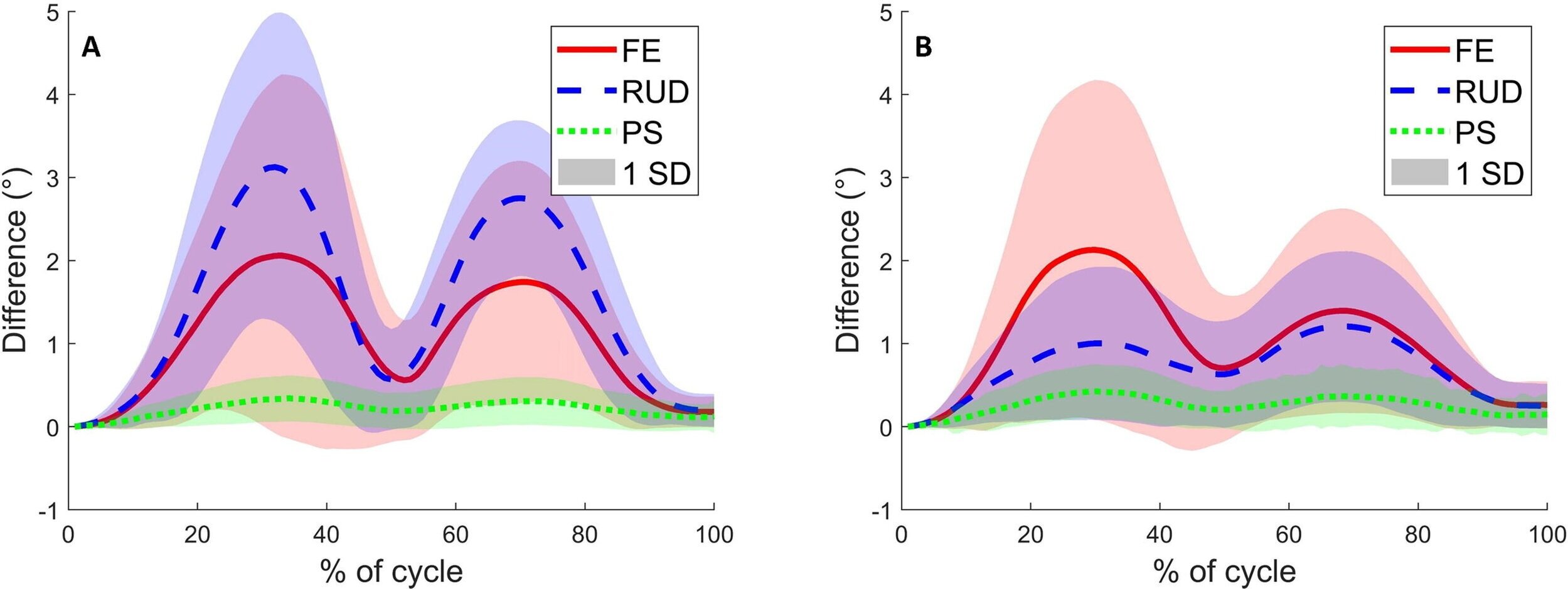
Mean ± one standard deviation (SD) of the kinematic differences in flexion–extension (FE), radioulnar deviation (RUD), and pronation-supination (PS) between the recommended joint coordinate system and the ISB joint coordinate system for all participants during (A) FE and (B) RUD.

Mean ± one standard deviation (SD) of the kinematic differences in flexion–extension (FE), radioulnar deviation (RUD), and pronation-supination (PS) resulting from two sets of digitisations built using (A & B) the ISB joint coordinate system and (C & D) the recommended joint coordinate system. The differences during nine repetitions were averaged for all 23 participants performing (A & C) FE and (B & D) RUD.
2020
Akinnola OO, Vardakastani V, Kedgley AE. (2020). The effect of planar constraint on the definition of the wrist axes of rotation. Journal of Biomechanics,113, 110083. doi: 10.1016/j.jbiomech.2020.110083
Akinnola OO, Vardakastani V, Kedgley AE. (2020). Identifying tasks to elicit maximum voluntary contraction in the muscles of the forearm. Journal of Electromyography and Kinesiology, 55, 102463. doi: 10.1016/j.jelekin.2020.102463
Shah DS, Middleton C, Gurdezi S, Horwitz MD, Kedgley AE. (2020). The Effect of Surgical Treatments for Trapeziometacarpal Osteoarthritis on Wrist Biomechanics: A Cadaver Study. The Journal of Hand Surgery, 45(5), 389–398. doi: 10.1016/j.jhsa.2019.10.003
Shah DS, Horwitz MD, Kedgley AE. (2020). Extensor retinaculum excision does not affect wrist tendon forces: a cadaveric simulator study. Journal of Hand Surgery (European Volume), 45(9), 986–988. doi: 10.1177/1753193420928781
Nolte D, Ko S-T, Bull AMJ, Kedgley AE. (2020) Reconstruction of the lower limb bones from digitised anatomical landmarks using statistical shape modelling. Gait & Posture, 77, 269–275. doi: 10.1016/j.gaitpost.2020.02.010
Jones D, Wang L, Ghanbari A, Vardakastani V, Kedgley AE, Gardiner MD, Vincent TL, Culmer PR, Alazmani A. (2020) Design and Evaluation of Magnetic Hall Effect Tactile Sensors for Use in Sensorized Splints. Sensors, 20(4), 1123. MDPI AG. doi: 0.3390/s20041123
Rusli WMR & Kedgley AE. (2020) Statistical shape modelling of the first carpometacarpal joint reveals high variation in morphology. Biomechanics and modeling in mechanobiology, 19(4), 1203–1210. doi: 10.1007/s10237-019-01257-8

A representative plot of the instantaneous helical axes (IHAs) (coloured axes) of the wrist during flexion-extension in the transverse (A), coronal (B), and sagittal (C) planes, as well as an isometric (D) view. The black vector is the screw displacement axis that is derived from the IHAs.

Diagrammatic representation of safe zone measurement; indicating the arc and perimeter measurements with respect to the ECU tendon and distal ulna articular surface. ECU = extensor carpi ulnaris; K-wire = kirschner wires.

Mean muscle forces of the APL, FCR, and ECU across 9 specimens during FE-5030 and RUD-15 in the intact specimens (dashed lines) and following trapeziectomy (solid lines). Error bars represent 1 SD. The asterisk (*) represents statistically significant differences between trapeziectomy and intact cases (P < .01).
prior to 2020
Carpanen D, Kedgley AE, Shah DS, Edwards DS, Plant DJ & Masouros SD. (2019). Injury risk of interphalangeal and metacarpophalangeal joints under impact loading. Journal of the Mechanical Behavior of Biomedical Materials, 97, 306–311. doi: 10.1016/j.jmbbm.2019.05.037
Ding Z, Tsang CK, Nolte D, Kedgley AE, & Bull AMJB. (2019) Improving Musculoskeletal Model Scaling Using an Anatomical Atlas: The Importance of Gender and Anthropometric Similarity to Quantify Joint Reaction Forces. IEEE transactions on bio-medical engineering, 66(12), 3444–3456. doi: 10.1109/TBME.2019.2905956
Kedgley AE, Saw T, Segal NA, Hansen UN, Bull AMJ, Masouros SD. (2019) Predicting meniscal tear stability across knee-joint flexion using finite-element analysis. Knee Surg Sports Traumatol Arthrosc 27, 206–214. doi: 0.1007/s00167-018-5090-4
Shah DS, Middleton C, Gurdezi S, Horwitz MD, Kedgley AE. (2018) Alterations to wrist tendon forces following flexor carpi radialis or ulnaris sacrifice: a cadaveric simulator study, Journal of Hand Surgery (European) 43: 886-888. doi: 10.1177/1753193418783176
Shah DS, Middleton C, Gurdezi S, Horwitz MD, Kedgley AE. (2018) The importance of abductor pollicis longus in wrist motions: A physiological wrist simulator study, Journal of Biomechanics 77: 218-222. doi: 10.1016/j.jbiomech.2018.07.011
Vardakastani V, Bell H, Mee S, Brigstocke G, Kedgley AE. (2018) Clinical measurement of the dart throwing motion of the wrist: variability, accuracy and correction, Journal of Hand Surgery (European) 43: 723-731. doi: 10.1177/1753193418773329
Garland AK, Shah DS, Kedgley AE. (2018) Wrist tendon moment arms: Quantification by imaging and experimental techniques, Journal of Biomechanics 68: 136-140. doi: 10.1016/j.jbiomech.2017.12.024
Goislard De Monsabert B, Edwards D, Shah DS, Kedgley AE. (2018) Importance of consistent datasets in musculoskeletal modelling: A study of the hand and wrist, Annals of Biomedical Engineering 46: 71–85. doi: 10.1007/s10439-017-1936-z
Taylor SAF, Kedgley AE, Humphries A, Shaheen AF. (2018) Simulated activities of daily living do not replicate functional upper limb movement or reduce movement variability, Journal of Biomechanics 76: 119-128. doi: 10.1016/j.jbiomech.2018.05.040
Shah DS, Middleton C, Gurdezi S, Horwitz MD, Kedgley AE. (2017) The effects of wrist motion and hand orientation on muscle forces: A physiologic wrist simulator study, Journal of Biomechanics 60: 232-237. doi: 10.1016/j.jbiomech.2017.06.017
Shah DS, Kedgley AE. (2016) Control of a wrist joint motion simulator: A phantom study, Journal of Biomechanics 49: 3061-3068. doi: 10.1016/j.jbiomech.2016.07.001
Amabile C, Bull AMJ, Kedgley AE. (2016) The centre of rotation of the shoulder complex and the effect of normalisation, Journal of Biomechanics 49: 1938-1943. doi: 10.1016/j.jbiomech.2016.03.035

Goniometry measurements. At the start of the motion: (a) extension, (b) DTM and (c) radial deviation angle. At the end of the motion: (d) flexion, (e) DTM and (f) ulnar deviation angle. For measurements of the DTM angle, the arms of goniometer were aligned with the dorsal side of the radius and the second metacarpal. For all other measurements, the goniometer was placed according to standard clinical practice.

A multivariate box-and-whisker plot of posterior radial displacement at the maximum load (Dmax, mm) at 0°, 30°, 60° and 90° of elbow flexion of the normal (control) elbow and after isolated Osborne-Cotterill lesion (OCL) and OCL + lateral collateral ligament complex (LCLC) resection (group 1). The horizontal line in the middle of each box indicates the median, the top and bottom borders of the box mark the 75th and 25th percentiles, respectively, and the whiskers indicate the standard deviation.

The maximum, minimum and ranges of motion for the thoracic lateral flexion, axial rotation and forward flexion for the eat, wash, retrieve from shelf, comb and perineal care ADLs. Simulated tasks (STs) are shown in solid grey and functional tasks (FTs) are shown in patterned grey, error bars represent ± one standard deviation of the means of maximum and minimum angles. * show significant differences in maximum/minimum angles and § show a significant difference in the range of motion.

Muscle forces (mean ± one standard deviation) in flexion–extension (FE-5030) with (dashed) and without (solid) the abductor pollicis longus (APL) for flexor carpi radialis (FCR), extensor carpi radialis longus (ECRL) and extensor carpi ulnaris (ECU). The asterisk (*) indicates statistically significant differences between the two groups (significance: p < 0.05).